What is the vestibulocochlear nerve?
The vestibulocochlear nerve (CN8) or the auditory vestibular nerve is the eighth pair of cranial nerves from twelve pairs. This nerve is comprised of two portions- both parts having sensory functions. This nerve is responsible for transmitting sound information as well as for maintaining balance and equilibrium, transmitting the information from the inner ear to the brain. A human beings sense of equilibrium is determined by the vestibulocochlear nerve.
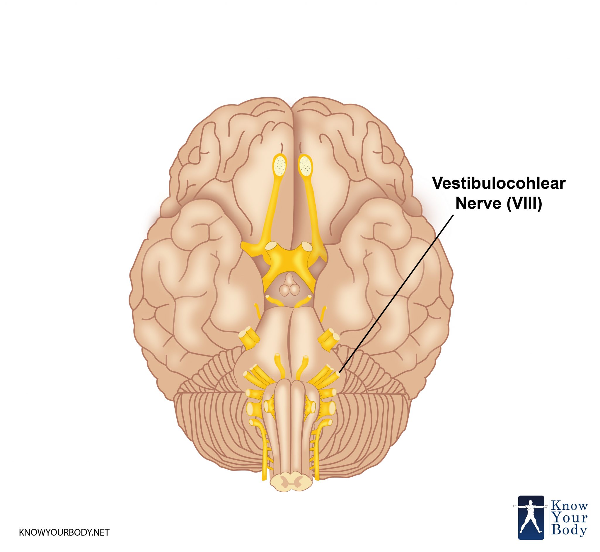
Vestibulocochlear Nerve Location
The vestibulocochlear nerves are the eighth pair of twelve cranial nerves. These sensory fibers consist of afferent sensory fibers, which means that the vestibulocochlear nerves towards the center; from the peripheral limbs towards the central nervous system.
There are two parts of the vestibulocochlear nerve- the vestibular component and the cochlear component.
• The Vestibular Component arises from the vestibular nuclei complex, arising from the pons and the medulla. This vestibule is filled with endolymphatic fluid, and it is responsible for the sense of somatic balance and equilibrium in a human being.
• The Cochlear Component arises from the ventral and dorsal cochlear nuclei, which are present in the inferior cerebellar peduncle. This nerve is responsible for the sensory hearing function of a human being.
Vestibulocochlear Nerve Location
Vestibulocochlear Nerve Function
The vestibulocochlear nerve has two special sensory functions which are carried out by the cochlear nerves and the vestibular nerves.
• Hearing Function: The cochlear nerves are responsible for detecting the frequency and the magnitude of sound vibrations and waves. The inner hair cells located inside the inner ear receive these sound waves and transmit the information from the inner ear back to the brain, where it is processed. The magnitude of the sound waves received by the inner ear is determined by how much the inner ear membrane vibrates, and thus how much the vibrations are triggered in the inner ear cells. The more the vibrations, the louder the sounds. The frequency of the sound waves is determined by the position of the activated inner hair cells along the membrane.
• Balance Function: The vestibular component of the vestibulocochlear nerves are responsible for sensing the minute changes in the position of the head, in relation to the gravity around us. The vestibular cells can detect linear as well as rotational movements of an individuals head. These cells are located inside the inner ear, in the otolith organs as well as in the semi-circular canals. Information is sent from the vestibular cells in the inner ear to the brain, and this information is used to coordinate balance in the human body. The Vestibule-ocular reflex is also determined by these cells. This Vestibulo-ocular reflex stabilizes the images which we project on our retina, by moving our eyes in the opposite direction to which we are turning our heads.
Vestibulocochlear Nerve Origin
The vestibulocochlear nerve consists of two portions; the vestibular portion and the cochlear portion. The vestibular portion of the vestibulocochlear nerve originates in the vestibular ganglion, which is a group of nerve cells located in the internal acoustic meatus. This is in the form of a channel in the temporal bone of the skull through which facial nerves, auditory nerves, and blood vessels run.
Vestibulocochlear Nerve Structure
The basic structure of the vestibulocochlear nerve consists of bipolar nerves- the vestibular nerve and the cochlear nerve.
The vestibular nerve travels from the inner portion of the ear. The vestibular ganglion is where the cell bodies of the bipolar neurons stay and extend their processes to the five sensory organs.
The cochlear nerve travels away from the cochlea of the inner ear. From here, it is seen to start in the form of spiral ganglia. The inner ear hair cells of the organ of Corti are responsible for the activation of the various receptors, which receive the pressure waves and respond to sound, sending information from the inner ear to the brain.
Vestibulocochlear Nerve Anatomy
The vestibulocochlear nerve consists of two sets of fiber branches; the vestibular nerve and the cochlear nerve. Both these nerves are anatomically as well as physiologically different.
The peripheral portions of both the nerves; the vestibular nerve and the cochlear nerve join at the lateral portion of the internal auditory canal. It is at this joining, that they form the vestibulocochlear nerve.
Vestibulocochlear Nerve Innervation
The vestibulocochlear nerve innervates the cochlear for hearing, and it also innervates the vestibule for acceleration sensing.
Related Conditions
Signs and Symptoms:
Damage to the vestibulocochlear nerves can cause symptoms such as:
• Motion sickness
• False sense of motion
• Hearing loss
• Vertigo
• Loss of equilibrium in dark places
• Gaze-evoked tinnitus
• nystagmus
Causes of damage:
Some of the causes of damage to the inner ear and vestibulocochlear nerves are:
1. Trauma, head injuries, injury to the ear, and skull fractures can cause damage to the vestibulocochlear nerves. This can result in permanent or semi-permanent hearing loss, hearing impairment as well as a loss a sense of balance.
2. Excessive noise and prolonged exposure, or even sudden exposures to very loud noises can cause hearing damage and hearing loss. This can be due to damage to the vestibulocochlear nerve endings, and can cause hearing loss or tinnitus, which is often referred to as the sensation of hearing a ‘ringing in the ears’.
3. Tumors on the skull, the brain, and the inner ear, could damage the acoustic and vestibular branches of the vestibulocochlear nerves. Hearing loss, balance disorders, damage to the hearing as well as tinnitus are some of the symptoms of a tumor which damages the vestibulocochlear nerves.
Types of vestibulocochlear nerve disorders:
1. Vestibular neuritis- This refers to the inflammation of the vestibular branch of the vestibulocochlear nerve. Some of the symptoms of this disorder are nausea and vomiting, loss of equilibrium especially in dark places, vertigo, and nystagmus.
2. Labyrinths- this disorder refers to an inflammation of the cochlea as well as the vestibular branches of the vestibulocochlear nerve. The symptoms presented are similar to those of vestibular neuritis, with the exception of tinnitus as well as sensorineural hearing loss.
3. Acoustic neuroma- the development of a serious, yet a non-malignant tumor in the inner ear cavity. As the tumor grows, it compresses the vestibulocochlear nerve, thus compromising the ability to hear, sense of balance, causes vertigo, sensory hearing loss, dizziness, nausea, etc.
4. Benign Paroxysmal Positional Vertigo (BPPV)- when excessive debris gets collected in the inner ear cavity, it causes BPPV, with symptoms such as vertigo, dizziness, and other vestibular disorder related symptoms.
5. A migraine associated vertigo or MAV is when a patient experiences symptoms of vertigo due to an excessive migraine. Patients can experience dizziness, vertigo, nausea, and loss of balance during migraine attacks.
6. Some of the other types of vestibular disorders are:
• Age-related dizziness and imbalance
• Autoimmune inner ear disease
• Bilateral vestibular hypofunction
• Canvas syndrome
• Cervicogenic dizziness
• Cholesteatoma
• Concussion
• Neurotoxicity
• Ototoxicity
Treatment for Vestibulocochlear Nerve
Depending on the type of vestibular disorder, the history of the patient, the extent of the injury and the disorder, there are several different types of treatment plans that can be pursued.
1. Vestibular rehabilitation therapy or VRT uses head, eye and body exercises to re-train the brain to process the signals from the vestibular system. Then, the brain can coordinate this information with the other parts of the body.
2. Home-based exercise performed by the patient on his own, or with the help of certified therapists can be extremely helpful in the treatment of vestibulocochlear nerve disorders.
3. Medication can also be helpful in treating the symptoms of vestibulocochlear nerve disorders
4. As a last resort, if none of the methods of treatment work on the patient, they can go through surgery to repair the symptoms of vertigo, dizziness and other symptoms related to vestibulocochlear nerve disorders.
Frequently Asked Questions
What is the purpose and function of the vestibulocochlear nerve?
The vestibulocochlear nerve is responsible for both the hearing function as well as the balance function in a human being. The vestibulocochlear nerve transmits information from the inner ear to the brain, which receives and processes this information. The cochlea and the vestibular apparatus allow the vestibulocochlear nerve to function properly.
What is an acoustic neuroma?
Also known as a vestibular schwannoma, the acoustic neuroma is a non-cancerous tumor growth. This tumor can often put pressure on the vestibulocochlear nerve in the inner ear, causing problems with a person’s hearing ability as well as ability to maintain balance and equilibrium.
What are the risk factors associated with vestibular schwannoma?
Partial or complete loss of hearing, loss of sense of acceleration and balance, dizziness, nausea associated with the symptoms.
No comments yet.